Nuclear Associates 76-908 & 76-907
Operators Manual
2-10
2.4 Tests With Uniformity and Linearity (UAL) Phantom
2.4.1 Image Uniformity
Image uniformity refers to the ability of the MR imaging system to produce a constant signal response
throughout the scanned volume when the object being imaged has homogeneous MR characteristics.
Factors Affecting Image Uniformity include:
(1) Static-field inhomogeneities
(2) RF field non-uniformity
(3) Eddy currents
(4) Gradient pulse calibration
(5) Image processing
Any typical multi-slice acquisition may be used provided the signal-to-noise ratio is sufficiently large so
that it does not affect the uniformity measurement. Adequate signal-to-noise may be insured by either
increasing the number of acquisitions (NEX) or by applying a low-pass smoothing filter. In practice, it has
been found that a signal-to-noise ratio of 80:1 or greater will yield good results.
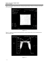
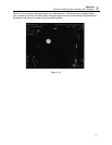
For pixels within a centered geometric area that encloses approximately 75% of the phantom area, the
maximum (S max) and minimum (S min) values are determined. Care should be taken to not include
edge artifacts in the ROI.
A range (R) and mid-range value S are calculated as follows:
R = (S max- S min) /2
S = (S max+ S min) / 2
The relationship for calculating integral uniformity (U) is:
U = (1 – (R/S)) x 100%
Perfect integral uniformity using this relationship is when U = 100%
In some cases (e.g., low-field imaging) signal-to-noise may be a limiting factor in the measurement of
image uniformity. To help minimize the effect of noise on the measurement the image may be convolved
with a 9-point low-pass filter.
For a 20 cm field-of-view or less, the uniformity should be typically 80% or better. It should be realized
that for larger fields-of-view, the uniformity may diminish. Image uniformity in the above context is not
defined for surface coils.
SNR results are only applicable to the specific system, phantom and scan conditions. It is important to re-
emphasize that the signal and noise measurements are dependent on essentially all scan parameters
and test conditions. SNR should be normalized to voxel size for comparison.
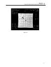
2.4.2 Spatial Linearity (Distortion)
Spatial linearity describes the degree of geometrical distortion present in images produced by any
imaging system. Geometrical distortion can refer to either displacement of displayed points within an
image relative to their known location, or improper scaling of the distance between points anywhere within
the image.
The primary factors that introduce geometrical distortion in MR imaging are:
(1) Inhomogeneity of the main magnetic field


