


11
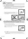
2 Preparation
¿
Turning OFF the instrument without following the above procedures may result in a loss of data or
damage to the instrument. With an exception of emergency situations, follow the above procedures to
turn OFF the instrument.
¿
The power switch does not go OFF automatically. Do not forget to turn OFF the power switch.
2.6 Retinal camera preparation
Following the daily inspection list in “7.2 Daily inspection”, prepare the retinal camera for operation.
2.7 Preparatory procedure of the examined eye
1
In case of mydriatic photography, apply mydriatic eye drops to the examined eye. After it dilates fully,
guide the patient to the retinal camera. In case of non-mydriatic photography, take the patient to a dark
room and let the examined eye dilate spontaneously.
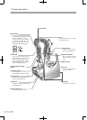
2
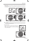
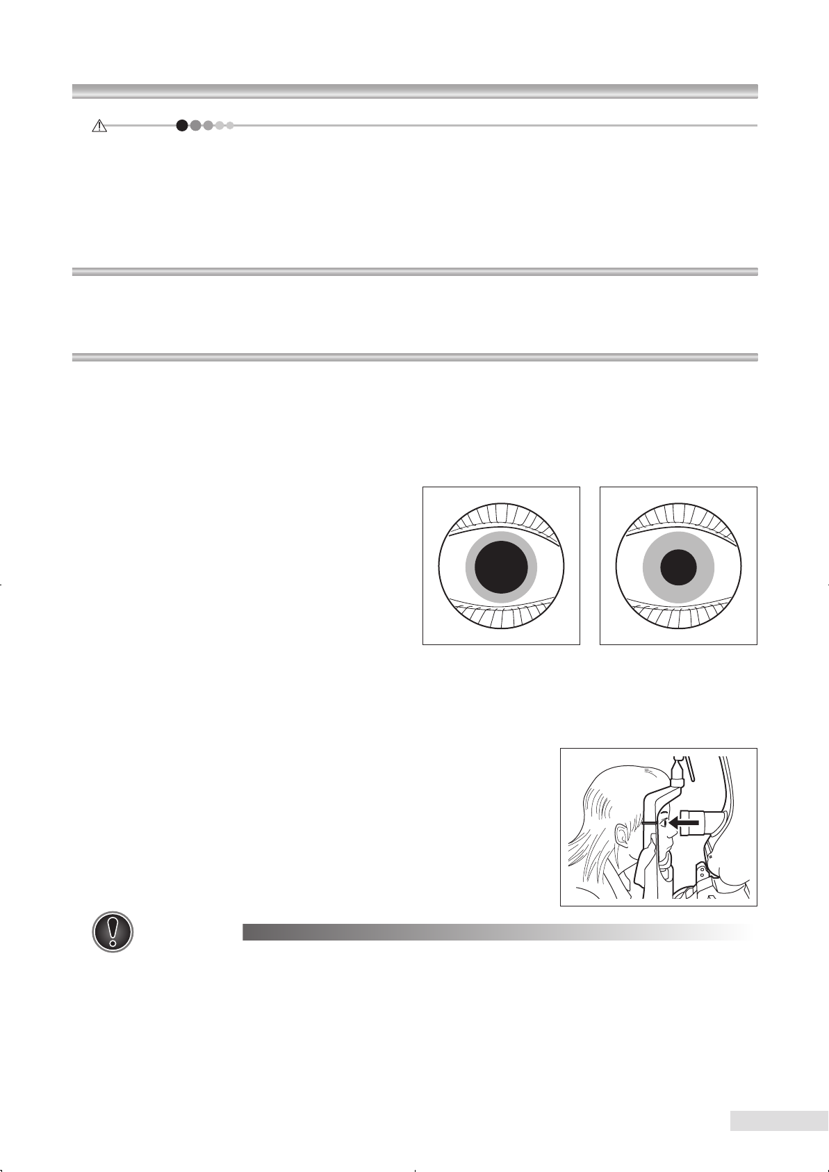
Make sure that the pupil is sufciently open.
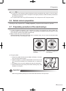
Sufcient diameter of the pupil is 5.5 mm or more in mydriatic mode, 4.0 mm or more in small pupil mode
and 4.0 mm or more in non-mydriatic mode.
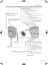
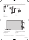
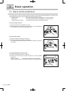
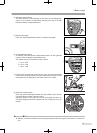
3) Fixing the patient
1. Instruct the patient whose eyes are dilated sufciently, to be seat-
ed in front of the retinal camera.
2. Adjust the height of a powered optical table to let the chin on the
chin rest and the forehead on the forehead rest in a natural pos-
ture.
3. The height of chin rest can be adjusted with the chin rest buttons.
4. Set the examined eye at the eye level mark indicating lamp. (See
illustration on the right.)
Important
◇ When using mydriatic eye drops, be sure to follow the instruction of the eye drops.
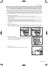
Mydriatic photography is
enabled if 5.5 mm or more
in diameter.
Small pupil or non-mydriatic
photography is enabled if 4.0
mm in diameter.